Extracellular Vesicles Analysis Services
Online InquiryExtracellular vesicles (EVs) are a general term for a variety of vesicles with lipid bilayer membrane structures released by cells at rest or under stress, and are important mediators of intercellular signaling common to eukaryotic and prokaryotic cells. Depending on how EVs are generated, their size or function, they can be divided into different subgroups such as exosomes, microvesicles, migratory bodies and apoptotic vesicles. The classification is mainly based on the morphological structure, size and volume of extracellular vesicles, their mode of occurrence and specific molecular markers. Extracellular vesicles can deliver proteins, lipids and nucleic acids that affect the physiological and pathophysiological functions of donor and recipient cells.
Extracellular vesicles are found in a variety of body fluids, including blood, urine, saliva, breast milk, amniotic fluid, cerebrospinal fluid, and synovial fluid. Detection of extracellular vesicles in body fluids can indicate the course of disease and facilitate clinical biomarker discovery. Creative Proteomics provides you with a professional platform for multi-group analysis of extracellular vesicles. We have high resolution mass spectrometry instruments and a professional bioinformatics team to provide you with extracellular vesicle proteomics, extracellular vesicle metabolomics and extracellular vesicle lipidomics analysis services. Our aim is to provide you with excellent services, accelerate the progress of your project and open up new horizons.
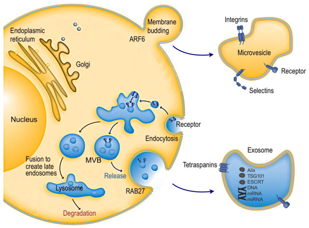
 Intracellular pathways of EV biogenesis and secretion (Shao et al., 2018)
The Extracellular Vesicles Assays We Offer Include but Are Not Limited to:
1. Extracellular vesicles isolation and purification
We have a standard process from collection of body fluid samples or in vitro cultured cell samples to sample analysis, with the option/combination of ultra-centrifugation or density gradient centrifugation, affinity purification, nanotechnology, microfluidic analysis and other methods to obtain purer extracellular vesicles.
2. Detection of morphology and purity of extracellular vesicles
- Electron microscope inspection
- Nanoparticle tracking analysis techniques
- Flow cytometry detection
3. Extracellular vesicles proteomics analysis
- Characterization of transmembrane proteins: exosomes are rich in adenosine tetraphosphate and microvesicles are rich in integrins, selectins and CD40 ligands. Many of the transmembrane proteins are involved in normal physiology and disease pathogenesis, therefore, studying transmembrane proteins may reveal important pathophysiological EV biomarkers.
- Intracapsular protein characterization: Characterization of intracapsular proteins such as cytoplasmic proteins (e.g. TSG101, ALIX, membrane associates and Rabs), cytoskeletal proteins (e.g. actin, myosin, microtubulin), molecular chaperone proteins (e.g. heat shock proteins/heat shock proteins), metabolic enzymes (e.g. enolase, glyceraldehyde 3-phosphate dehydrogenase/GAPDH) and ribosomal proteins.
- Unknown protein identification
- Full spectrum analysis of extracellular vesicular proteins
- Differential expression analysis of extracellular vesicular proteins
- Protein-protein interaction analysis
4. Extracellular vesicles metabolomics analysis
- Extracellular vesicles metabolite differential expression analysis
- Qualitative and quantitative analysis of extracellular vesicles metabolites
5. Extracellular vesicles lipidomic analysis
- Fatty acid profiling of extracellular vesicles
- Qualitative and quantitative analysis of lipid
- Differential analysis of extracellular vesicle lipid molecule levels
Basic Analysis
- Data quality control and molecular identification
- Expression quantitative analysis
- Differential analysis
- Functional annotation and enrichment analysis
Reference
- Shao, H., Im, H., et al. (2018). New technologies for analysis of extracellular vesicles. Chemical reviews, 118(4), 1917-1950.
* For Research Use Only. Not for use in diagnostic procedures.


