Exosomal Biogenesis and Identification
Online InquiryAs a subset of extracellular vesicles (EVs), initially, exosomes were thought to be a cellular mechanism for the excretion of unwanted cellular products. Exosomes transfer functional proteins, nucleic acids (such as miRNA and mRNA), and metabolites to recipient cells, playing significant roles in cellular communication and influencing a broad range of physiological and pathological processes. Although exosomes have been extensively studied, the biogenesis of exosomes is complex and requires further research. Creative Proteomics is a leading custom service provider in exosome analysis. We can provide you with exosome proteomics, exosome metabolomics and exosome lipidomics analysis services, but are not limited to them. Our capabilities will provide strong support for the study of exosome biogenesis and its identification.
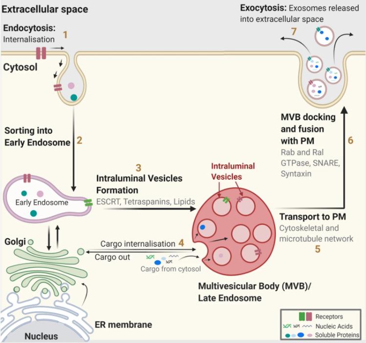 Fig. 1 Exosome biogenesis. (Gurung, Sonam, et
al., 2021)
Exosome biogenesis solution
In the endosomal system, internalized cargoes are sorted into early endosomes, which then mature into multivesicular bodies (MVBs) or late endosomes. MVBs are transported to the plasma membrane through the cytoskeleton and microtubule network, fused to the cell surface and released intraluminal vesicles (ILVs). ILVs contained in MVBs and late endosomes are the precursors of exosomes. Moreover, several other degradation pathways exist for MVBs, and it is unclear whether the secretion and degradation pathways of MVBs are different. To date, there are multiple mechanisms involved in exosome biogenesis and secretion. ESCRT machinery (ESCRT proteins) is a cytoplasmic multi-subunit system essential for membrane remodeling, which plays a prominent role in ILV biogenesis. In addition to ESCRT-dependent processes, the role of complex lipids and other protein-related pathways (such as tetraspanins, Rab GTPases, and autophagy-related proteins) in exosome production cannot be underestimated.
Creative Proteomics has over a decade of experience in multi-omics services. With our deep expertise and advanced technology, we will help you obtain single-cell and subcellular-level views to facilitate relevant research progress. We have the ability to help our global customers improve their understanding of exosome production and release kinetics.
Exosome heterogeneity and identification
Exosomes are a heterogeneous population due to their differences in cellular origin, size, composition, and function. Thus, the identification and analysis of exosomes are more complex. Exosomes can be differentiated and classed by their sizes and density. It is believed that the differences in exosomes are related to the limiting membrane of MVBs during ILV formation and the molecular pathways of exosome biogenesis. In addition to differences in their biophysical properties and composition, heterogeneity can lead to different exosome contents.
Recognizing the heterogeneity of exosomes is important to determine the content of cargoes, the functional role, and allow for better exosome analysis. Creative Proteomics offers the most popular isolation methods for exosome generation and identification such as ultracentrifugation and size exclusion coupled with analysis methods such as western blotting (WB), nanoparticle tracking analysis (NTA), and transmission electron microscopy (TEM). In addition, we have adopted a global and targeted proteomics and lipidomics approach to provide further assistance in related studies.
Advantages of Creative Proteomics
- Advanced technology platform
- Get better data on exosomes
- Standardized workflow and strict quality control
- Full technical tracking service and guidance
If you have a question or want general information, please feel free to contact us.
References
- Gurung, Sonam, et al. "The exosome journey: From biogenesis to uptake and intracellular signalling." Cell Communication and Signaling 19.1 (2021): 1-19.
- Kalluri, Raghu, and Valerie S. LeBleu. "The biology, function, and biomedical applications of exosomes." Science 367.6478 (2020): eaau6977.
Related Services
* For Research Use Only. Not for use in diagnostic procedures.


