Journal: Oncotarget
Published: 2018
Background
This study explores how the inhibition of the mevalonate pathway, particularly through statin drugs like lovastatin, influences metabolic and lipidomic profiles in cancer cells, specifically HGT-1 human gastric cancer cells. Previous research has shown that combining statins with taxane drugs like docetaxel promotes apoptosis in these cells by disrupting cholesterol synthesis and post-translational prenylation, critical for the function of Ras and Rho proteins involved in growth signaling. Using a multi-platform approach integrating mass spectrometry (GC-MS, LC-MS) and nuclear magnetic resonance (NMR), this study identified over 100 metabolites with altered levels following treatment, emphasizing the potential of combining metabolomics, lipidomics, and transcriptomics to discover biomarkers for cancer cell apoptosis.
Materials & Methods
Cell Culture and Treatments
HGT-1 gastric cancer cells, along with AGS (gastric) and HCT116 (colon) cells, were grown in DMEM with 4.5 g/L glucose and 5% fetal bovine serum. Cells were treated at ~80% confluence with lovastatin (12.5 μM) and/or docetaxel (5 nM) for up to 36 hours, using DMSO as a vehicle control. Lovastatin treatment induced apoptosis at 36 hours, as indicated by morphological changes. Cells were then lysed in methanol for metabolite extraction.
HMG-CoA Reductase Activity
To measure HMG-CoA reductase activity, cell lysates were prepared and incubated with [14C]HMG-CoA and NADPH, followed by HPLC analysis. Enzyme activity was calculated as pmol/min/mg protein.
Metabolomics and Lipidomics Analysis
Samples were analyzed across five platforms: NMR (Bruker), GC-MS (Agilent), and three LC-MS systems (Thermo and Waters). Quality control (QC) samples helped correct analytical drift, with lipid markers having <30% RSD in QC samples retained. Key lipid and metabolite markers, including glutamine, were quantified by GC-MS and LC-MS.
Data Processing
Data normalization was performed using log10 and Pareto transformations. Principal component analysis (PCA) assessed treatment effects, with the RV coefficient verifying consistency across datasets. All analyses were conducted in FactoMineR and Workflow4Metabolomics.
Results
Inhibition of HMG-CoA Reductase
Lovastatin, alone or in combination with docetaxel, suppressed HMG-CoA reductase activity, leading to a full halt in mevalonate production, as evidenced by the basal enzyme activity dropping from 18 ± 2 pmol/min/mg protein to undetectable levels. Docetaxel alone had no measurable impact on HMG-CoA reductase activity.
Principal Component Analysis (PCA)
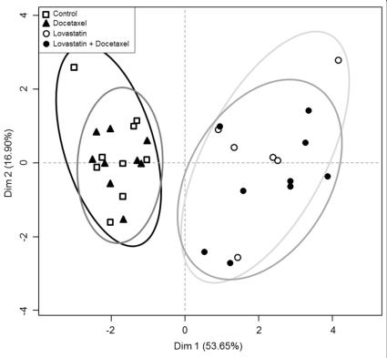
PCA revealed clear separations between lovastatin-treated and non-lovastatin-treated samples, with the former showing strong metabolomic and lipidomic shifts across HGT-1 gastric, AGS gastric, and HCT116 colon cancer cell lines. Docetaxel's effects were less pronounced, reflecting its minimal impact on gene expression.
Cross-Platform Data Standardization and Biomarker Identification
A total of 111 metabolites and lipids were identified as significantly altered across treatment conditions. Glutamine was highly induced by lovastatin and the combination treatment, while spermidine levels decreased. Four metabolite classes—ceramides, phosphatidylcholines, triglycerides, and sphingomyelins—were notably distinct in the (lovastatin + docetaxel) group. Biomarkers identified by multiple platforms included glutamine, glutamate, myo-inositol, creatine, lactic acid, and fumarate.
Kinetic Metabolomic Analysis
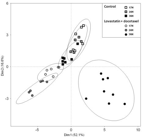
The kinetic analysis showed progressive changes in metabolite levels, with increases across most metabolites at 36 hours in the (lovastatin + docetaxel) group. Fumarate and pantothenic acid levels decreased consistently across all time points.
Integration of Metabolomics, Lipidomics, and Transcriptomics
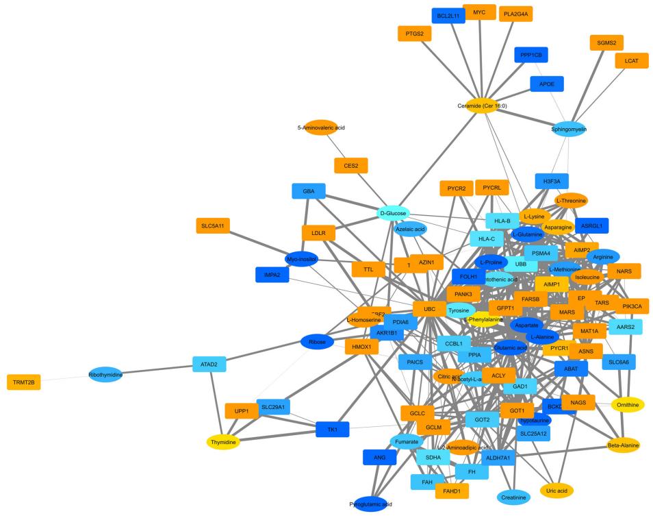
Data integration highlighted multiple upregulated genes in central carbon metabolism, including ACLY, ERBB2, and MYC, with corresponding increases in amino acids, citrate, lactate, fumarate, and other key metabolites. Downregulation of genes like P53, IDH, and MDH1 was observed, suggesting a reduction in P53-dependent apoptosis signaling and activation of MAP kinase pathways through ERBB2. The pathway analysis indicated that lovastatin treatments boost glucose uptake, amino acid synthesis, and lactate production, potentially increasing metabolic adaptations for cancer cell survival.
 Score plot of the "meta" PCA prepared from the calculated Dim1 coordinate from primary PCA analyses resulting from each data matrix.
 Combined network analysis of metabolites and lipids.
 PCA analysis of the kinetic effects of the (lovastatin + docetaxel) combination.
Reference
- Goulitquer, Sophie, et al. "Consequences of blunting the mevalonate pathway in cancer identified by a pluri-omics approach." Cell Death & Disease 9.7 (2018): 745.


