The mass spectrometer consists of three main components: the ion source, the mass analyzer, and the detector. In ion source, a sample is ionized, usually to cations by loss of an electron. And in the mass analyzer, the ions are sorted and separated according to their mass and charge. And in the detector, the separated ions are detected and relative abundance is recorded. Furthermore, the sample introduction system is essential to admin the samples into the ion source. And a computer and software are needed to control the instrument, obtain data and compare spectra according to the database.
The mass analyzer is the heart of the mass spectrometer, which takes ionized masses and separates them based on mass to charge ratios. There are several general types of mass analyzers, including magnetic sector, time of flight, quadrupole, ion trap.
1. Magnetic sector mass analyzer
In Magnetic Sector Mass Analyzer, ions are accelerated so that they have the same kinetic energy. All the ions are accelerated into a focused beam. And then the ions are deflected by the magnetic field according to masses of ions. The lighter ions have more deflection than the heavier ones. The amount of deflection depends on the number of the positive charges. When similar ions pass through the magnetic field, they all will be deflected to the same degree and will all follow the same trajectory path. Those ions which are not selected will collide with either side of the flight tube wall or will not pass through the slit to the detector.
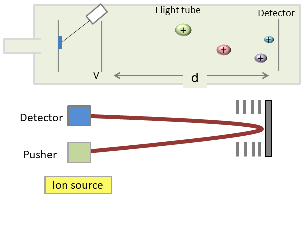
A time of flight analyzer consists of a pulsed ion source, an accelerating grid, a field-free flight tube, and a detector. The flight time needed by the ions with a particular mass to charge, accelerated by a potential voltage, to reach the detector placed at a distance, can be calculated from a formula.
Pulsing of the ion source is required to avoid the simultaneous arrival of ions of different m/z at the detector.
At high masses, not all the ions of the same m/z values reach their ideal velocities. To fix this problem, often a reflection which consists of a series of ring electrodes with high voltage is added to the end of the flight tube. Because of the high voltage, an ion is reflected in the opposite direction, when it into the reflection. For the ions of same m/z value, faster ions travel further than the slower ones into the reflections. In this way, both the slow and fast ions of the same m/z value reach the detector at the same time. The reflection increases resolution by narrowing the broadband range of flight times for a single m/z value.
3. Quadrupole

The quadrupole consists of four parallel metal rods and each opposing rod pair is connected together electrically. One pair of raids is applied with a radio frequency (RF) voltage while another one is applied with a direct current (DC) voltage. At a given DC and RF combination, only the ions of a particular m/z show a stable trajectory and can be transmitted to the detector, while other ions with unstable trajectories don’t pass the road, because the amplitude of their oscillation becomes infinite. By changing DC and RF in time, usually at a fixed ratio, ions with different m/z values can be transmitted to the detector one after another.
4. Ion trap
An ion trap is a device that uses an oscillating electric field to store ions. There are several common types of ion trap: 3D ion trap, linear ion trap, orbitrap and Fourier transform ion cyclotron resonance.
4.1 Quadrupole ion trap (QIT)
Quadrupole ion trap is also called 3 dimension ion trap. The QIT mass spectrometer uses three electrodes to trap ions in a small volume. It consists of a cylindrical ring electrode and two end-cap electrodes. A mass spectrum is obtained by changing the electrode voltages to eject the ions from the trap. The end-cap electrodes contain holes for the introduction of ions from an external ion source and for the ejection of ions toward an external detector.
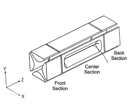
By creating a potential well for the ions, the linear ion trap can be used as a mass filter or as a trap. The linear ion trap uses a set of quadrupole rods to confine ions radially by a two-dimensional radio frequency (RF) field. And a static electrical potential can confine the ions axially. They are confined by application of appropriate RF and DC voltages with their final position maintained within the center section of the ion trap. The RF voltage is adjusted and multi-frequency resonance ejection waveforms are applied to the trap to eliminate all but the desired ions in preparation for subsequent fragmentation and mass analysis.
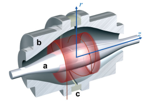
Orbitrap is the newest addition to the family of high-resolution mass analyzer. In orbitrap, moving ions are trapped in an electrostatic field. The electrostatic attraction towards the central electrode is compensated by a centrifugal force that arises from the initial tangential velocity of ions. The electrostatic field which ions experience inside the orbitrap forces them to move in complex spiral patterns. The axial component of these oscillations is independent of initial energy, angles and positions, and can be detected as an image current on the two halves of an electrode encapsulating the orbitrap. A Fourier transform is employed to obtain oscillation frequencies for ions with different masses, resulting in an accurate reading of their m/z.
4.4 The Fourier Transform Ion Cyclotron Resonance (FT-ICR)

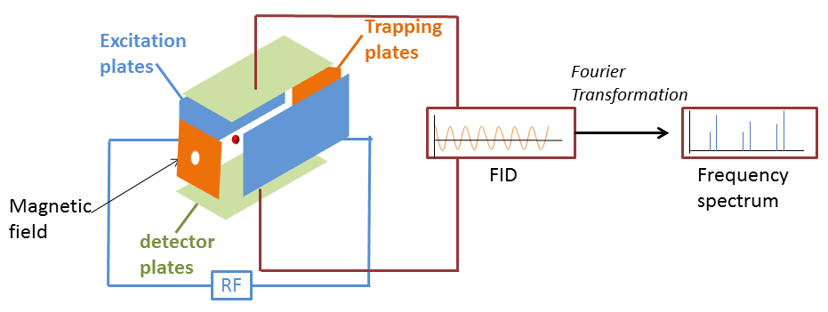
The Fourier Transform Ion Cyclotron Resonance mass spectrometer consists of three main sections: excitation plates, trapping plates, detector plats, and they consist a cell.
Ions which are affected by a magnetic field move at a cyclotron frequency. After a radio frequency voltage at the same frequency of cyclotron frequency is applied, the ions absorb energy and accelerate to a larger orbit radius than their original path. After excitation, the cyclotron radius of ions still remains the larger state. And as the ions around approach to the top and bottom plate, the electrons travel from top to bottom. The motion of electrons between these two plates produces a detectable current. The decay over time of the image current resulting after applying a short radio-frequency sweep is transformed from the time domain into a frequency domain signal by a Fourier transform.
References
1. Schwartz J C, Senko M W, Syka J E P. A two-dimensional quadrupole ion trap mass spectrometer. Journal of the American Society for Mass Spectrometry, 2002, 13(6): 659-669.
2. Scigelova M, Makarov A. Orbitrap mass analyzer–overview and applications in proteomics. Proteomics, 2006, 6(S2): 16-21.


