The Molecular Symphony: Phospholipid Structure
Hydrophilic Head: The hydrophilic head of a phospholipid is a complex but crucial component. It consists of a phosphate group (PO4) and a glycerol molecule. The phosphate group carries a negative charge, making it highly polar and attracted to water molecules. This polar region is vital for the interactions of phospholipids with the aqueous environment both inside and outside the cell. It acts as a "water-attracting" or hydrophilic component.
Hydrophobic Tails: The hydrophobic tails of phospholipids are long hydrocarbon chains. These chains are composed of carbon and hydrogen atoms and are nonpolar. Because of their nonpolar nature, they are repelled by water and tend to cluster together. This clustering is driven by the hydrophobic effect, a fundamental principle in chemistry, which seeks to minimize the contact between nonpolar substances and water. As a result, the hydrophobic tails align closely with each other, forming a hydrophobic core within the lipid bilayer.
Bilayer Formation: One of the most remarkable features of phospholipids is their ability to self-assemble into a bilayer structure. In this arrangement, phospholipids organize themselves so that their hydrophilic heads face outward, interacting with the surrounding aqueous environment (both inside and outside the cell), while their hydrophobic tails cluster inward, avoiding contact with water. This bilayer formation is the foundation of all cell membranes and is essential for creating a semi-permeable barrier that separates the inside of the cell from its external surroundings.
Architects of Life: Phospholipids and Cell Membranes
Cell membranes, often referred to as lipid bilayers, are the gatekeepers of cellular life. They owe their structural integrity and dynamic functionality to the presence of phospholipids. Let's delve into the intricate details of this critical relationship:
Lipid Bilayer Composition: The cell membrane consists primarily of a lipid bilayer, a double layer of phospholipid molecules. These phospholipids arrange themselves with their hydrophilic heads facing outward and their hydrophobic tails oriented inward. This arrangement creates a semi-permeable barrier that separates the interior of the cell from its external environment.
Selective Permeability: The lipid bilayer, composed mainly of hydrophobic tails, forms a formidable barrier to the passage of polar or charged molecules like ions, sugars, and amino acids. However, it allows the relatively small and nonpolar molecules such as oxygen, carbon dioxide, and lipids to pass freely. This selective permeability is essential for maintaining the internal environment of the cell.
Fluid Mosaic Model: The cell membrane is not a static structure but a dynamic mosaic. The fluid mosaic model describes it as a dynamic assembly of various molecules, including not only phospholipids but also integral and peripheral proteins, cholesterol molecules, and carbohydrates. These components can move laterally within the lipid bilayer, contributing to the membrane's flexibility and adaptability.
Cholesterol's Role: Cholesterol molecules are interspersed among the phospholipids in the lipid bilayer. Cholesterol acts as a "fluidity buffer." It makes the membrane less fluid at high temperatures by restricting the movement of phospholipids and more fluid at low temperatures by preventing close packing of phospholipids. This regulation of membrane fluidity is crucial for maintaining the membrane's integrity and functionality under varying environmental conditions.
Integral Membrane Proteins: Embedded within the lipid bilayer are integral membrane proteins. These proteins have hydrophobic regions that interact with the hydrophobic core of the bilayer. They play a variety of roles, such as serving as receptors for signaling molecules, transporters of ions and molecules, and enzymes that facilitate chemical reactions at the membrane.
Peripheral Proteins and Carbohydrates: Along the outer surface of the cell membrane, peripheral proteins and carbohydrates are found. Carbohydrates are often attached to proteins, forming glycoproteins, or to lipids, forming glycolipids. These structures are crucial for cell recognition, adhesion, and signaling.
Cell Membrane Flexibility: The dynamic nature of the cell membrane allows cells to change shape, fuse with other cells during processes like fertilization or immune responses, and carry out endocytosis and exocytosis. Phospholipids' ability to shift and adjust within the bilayer underpins these vital cellular functions.
Phospholipid Analysis Method
Sample Preparation: To begin, collect the biological sample of interest, which could be cell membranes, tissues, or biological fluids like blood or serum. Extract the lipids from the sample using a suitable solvent, typically a mixture of chloroform and methanol. After extraction, the solvent is evaporated to create a lipid film.
Phospholipid Extraction: To specifically analyze phospholipids, a phospholipid-specific extraction method is employed, such as the Bligh and Dyer method or the Folch method. These methods separate phospholipids from other lipid classes.
Phospholipid Hydrolysis: Hydrolyze the extracted phospholipids to release their constituent fatty acids. This is often achieved using an enzyme like phospholipase A2.
Derivatization: The liberated fatty acids may undergo derivatization, typically esterification, to make them suitable for analysis by gas chromatography (GC) or liquid chromatography (LC).
Chromatographic Separation: Chromatography, either GC or LC, is used to separate the derivatized phospholipid components based on their chemical properties. In GC, separation is achieved through vaporization and partitioning, while in LC, it relies on interactions between the stationary and mobile phases.
Detection: A suitable detector is employed based on the chromatographic system. For GC, this might be a flame ionization detector (FID), while for LC, it's often a mass spectrometer (MS). MS detection is highly sensitive and allows for the identification and quantification of individual phospholipid species.
Data Analysis: Analyze the chromatographic data to identify and quantify the various phospholipid species present in the sample. Software is used for peak integration and quantitation, and a phospholipid profile is generated, showing the types and relative concentrations of different phospholipids.

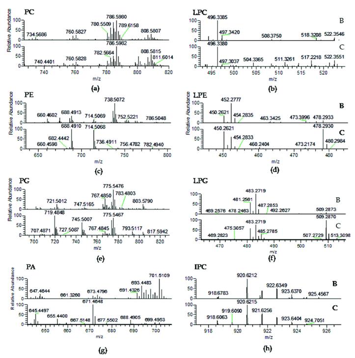
LC-MS spectra of phospholipid classes (Da et al., 2018)
Quantification: Quantify the phospholipids using calibration standards and software for accurate measurement.
Interpretation: Interpret the results within the context of the research objectives, which may include understanding lipid metabolism, membrane composition, or lipid alterations in disease states.
Reporting: Present the phospholipid analysis results in research papers, reports, or presentations, including the identified phospholipid species and their relative concentrations.
Reference
Da Costa, Elisabete, et al. "High-resolution lipidomics of the early life stages of the red seaweed Porphyra dioica." Molecules 23.1 (2018): 187.
